At Dentistry of Sugar Land, we rely on advanced imaging to make confident, well-informed treatment decisions. Cone-beam computed tomography (CBCT) gives our team high-resolution, three-dimensional views of teeth, bone, and surrounding anatomy so we can see what conventional X-rays sometimes miss. That clarity helps us diagnose more accurately and plan treatments with greater precision.
CBCT is a specialized tool designed for dental and maxillofacial care. It captures detailed volumetric data in a single, quick scan and presents information in multiple planes and slices. Used responsibly, CBCT enhances safety, shortens treatment timelines, and improves predictable outcomes across a broad range of procedures.
Traditional two-dimensional X-rays compress complex anatomy into a flat image, which can hide critical relationships between structures. CBCT restores depth, allowing clinicians to evaluate the exact position of roots, the shape and thickness of the jawbone, and the spatial relationship of teeth to nerves and sinuses. This three-dimensional context reduces guesswork when assessing complicated cases.
For endodontic concerns, CBCT can reveal hidden canals, vertical root fractures, or the true extent of resorption that conventional films may overlook. In oral pathology, the modality helps characterize lesions and determine whether additional testing or referral is warranted. In short, CBCT provides a clearer, more complete picture of the problem before treatment begins.
That clarity translates into better decisions for patients: fewer surprises during procedures, more targeted interventions, and a reduced likelihood of unnecessary treatment. When a situation is complex, CBCT is an invaluable diagnostic step that supports effective, conservative care.
Successful implant placement depends on understanding bone volume, density, and the exact location of vital structures. CBCT supplies accurate measurements in three planes so implant positions can be planned digitally and, when appropriate, transferred to a surgical guide. This level of planning supports more predictable implant placement and reduces intraoperative adjustments.
Similarly, for impacted teeth or complicated extractions, CBCT helps identify root angulations and proximity to nerves and sinuses. That information guides surgical approach and instrument selection, which can shorten procedure times and minimize tissue trauma. For restorations that interact closely with surrounding anatomy, three-dimensional imaging ensures restorations are designed with precise anatomic fit in mind.
In combination with digital impressions and CAD/CAM workflows, CBCT data becomes part of an integrated treatment plan that improves accuracy from diagnosis through final restoration. This coordinated approach benefits both clinical outcomes and patient comfort.
Although CBCT captures more anatomical detail than routine X-rays, modern units are optimized for dental use and permit targeted scanning of specific areas. Field-of-view options let clinicians limit exposure to the smallest region necessary for the diagnostic task, which helps keep radiation within recommended levels for dental imaging.
Our practice follows established guidelines for when CBCT is appropriate, balancing diagnostic benefit against exposure. The device’s quick acquisition time also reduces the chance of motion artifacts and repeated scans, further lowering cumulative dose. When CBCT is clinically justified, it provides essential information that outweighs the minimal additional exposure.
Beyond dose considerations, current CBCT systems provide sharp images with software tools that enhance visualization while preserving patient safety. These advancements allow clinicians to make confident choices without compromising on responsible imaging practices.
One of the practical benefits of CBCT is how it streamlines clinical workflows. Digital CBCT data can be viewed immediately, reformatted into cross-sections or panoramic reconstructions, and shared with specialists when collaborative care is needed. That immediacy shortens the diagnostic phase and helps coordinate multi-disciplinary treatment efficiently.
For patients, three-dimensional images are a powerful communication tool. Visualizing anatomy in 3D makes it easier to explain diagnoses and available options, which encourages informed decision-making. When patients can see the exact problem and the proposed plan, they typically feel more comfortable about next steps.
For referring clinicians and laboratory partners, CBCT files provide precise information for surgical guides, prosthetic design, and coordinated care. Clear data exchange reduces back-and-forth, supports accurate lab work, and helps keep treatment timelines on track.
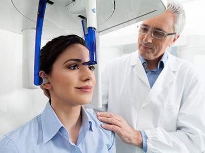
Preparing for a CBCT scan is straightforward. In most cases there is no special preparation: you’ll be asked to remove metal objects near the head and seated or positioned briefly in the scanner. The scan itself typically takes under a minute of actual imaging time, and patients often describe the experience as quick and comfortable.
After scanning, the imaging data are reconstructed into multiple views that your clinician reviews alongside clinical findings. Your dentist will explain the results in plain language, show relevant images, and outline how the information influences any recommended treatment. If a specialist’s input is needed, the digital files can be securely shared for additional evaluation.
Because CBCT is used selectively and purposefully, you can expect the scan to be part of a broader diagnostic plan rather than an exploratory step. When ordered, it adds clarity to the evaluation and helps the team design care that is precise, efficient, and tailored to your needs.
In summary, CBCT is a powerful diagnostic tool that gives clinicians the three-dimensional detail needed for confident decision-making, precise treatment planning, and improved communication. If you have questions about whether CBCT is appropriate for your situation or would like to learn more about how it might inform your care, please contact us for more information.


Cone-beam computed tomography (CBCT) is a three-dimensional imaging modality that captures volumetric views of the teeth, jaws, and surrounding facial structures. It acquires high-resolution data in a single, rapid scan and reconstructs images in axial, coronal, and sagittal planes. This volumetric detail allows clinicians to evaluate anatomy with depth and spatial accuracy not possible on standard two-dimensional films.
Unlike conventional dental X-rays, which compress anatomy into a flat image, CBCT preserves spatial relationships so clinicians can see root positions, bone contours, and the proximity of nerves and sinuses. That clarity improves diagnostic confidence for complex cases such as implant planning, impacted teeth, and endodontic evaluation. At Dentistry of Sugar Land, CBCT is used selectively to complement clinical examination and conventional radiography when additional detail is needed.
Clinicians recommend CBCT when two-dimensional images and clinical exam do not provide sufficient information to plan treatment safely and predictably. Common indications include implant site assessment, evaluation of impacted or ectopic teeth, complex endodontic cases, suspected lesions of the jaws, and airway or TMJ assessment. It is also useful for pre-surgical mapping in cases where vital structures lie close to the surgical field.
Each scan is ordered with a specific diagnostic question in mind rather than as a general screening tool. When CBCT is appropriate, the clinician selects the smallest field of view and imaging parameters necessary to answer the question, which limits exposure while capturing relevant detail. Scans are interpreted with clinical findings to form a comprehensive diagnosis and treatment plan.
For implants, CBCT provides precise measurements of bone height, width, and density while showing the location of adjacent teeth, nerves, and sinuses. These data allow for prosthetic-driven planning and virtual implant placement in three dimensions before the surgical appointment. The ability to plan digitally reduces unexpected intraoperative adjustments and supports fabrication of surgical guides when indicated.
CBCT also aids assessment of ridge defects, the need for grafting, and optimal implant angulation to achieve both functional and aesthetic outcomes. When combined with intraoral digital impressions and CAD/CAM workflows, CBCT datasets help ensure the final restoration fits accurately with the surrounding anatomy. This integrated approach improves predictability and can shorten overall treatment time.
Modern dental CBCT units are engineered for oral and maxillofacial imaging and offer adjustable fields of view and exposure settings. While CBCT delivers more anatomical detail than routine periapical or panoramic films, it generally exposes patients to less radiation than a conventional medical CT of the head. Clinicians apply the ALARA principle and only recommend scans when the diagnostic benefit outweighs the incremental exposure.
Targeted scans focused on a small region and rapid acquisition times reduce the likelihood of repeat imaging and lower cumulative dose. Our team follows established safety protocols, positions patients carefully, and uses shielding where appropriate to maintain responsible dosing. When clinically justified, the information gained from CBCT supports safer, more precise treatment.
Preparing for a CBCT scan usually requires no special medical preparation; patients are asked to remove jewelry, glasses, and removable dental appliances near the head. The patient will be seated or standing in the scanner and asked to remain still for the brief acquisition, which typically takes less than a minute. Many patients describe the procedure as quick and comfortably tolerated.
After acquisition, the volumetric data are reconstructed into multiple views that the clinician reviews alongside the clinical exam. Your dentist will explain the findings in plain language, point out relevant images, and describe how the information affects any recommended treatment. If specialist consultation is needed, digital files can be shared securely for coordinated care.
In endodontics, CBCT can reveal complex root canal anatomy, accessory canals, and curvatures that may be hidden on two-dimensional films. It is especially valuable for detecting vertical root fractures, the extent of internal or external resorption, and periapical pathology. This enhanced visualization can change case strategy, help identify retreatment needs, or indicate when surgical intervention is appropriate.
CBCT images allow clinicians to view teeth and surrounding bone in cross-section, which improves localization of lesions and assessment of their relationship to adjacent structures. That spatial information supports more conservative, targeted approaches and decreases the chance of unanticipated intraoperative findings. Interpretation requires experience and is always correlated with clinical signs and symptoms.
CBCT is not a replacement for conventional dental radiography in routine care; two-dimensional X-rays remain the standard for screening, routine caries detection, and periodic monitoring due to their low dose and proven effectiveness. CBCT is reserved for situations where three-dimensional detail will directly affect diagnosis or treatment planning. Using each modality appropriately ensures patients receive the most informative imaging with the least necessary exposure.
Dentists consider the clinical question, the alternatives, and applicable guidelines before ordering a CBCT scan. Where a CBCT would not materially change management, clinicians rely on traditional radiographs and clinical examination. This selective approach supports patient safety and diagnostic efficiency.
CBCT data are highly useful for communication because images can be reformatted into axial, sagittal, coronal, and three-dimensional renderings that patients and specialists can easily interpret. Showing a patient a cross-section or a 3D reconstruction helps translate clinical findings into understandable visual information and supports shared decision-making. For clinicians, the ability to export DICOM files enables seamless collaboration with oral surgeons, endodontists, and laboratories.
When appropriate, CBCT volumes are used to design surgical guides, plan implant positioning, and assist laboratory fabrication of restorations. Specialists reviewing the same datasets can provide coordinated recommendations that align with the primary clinician’s plan. This interoperability improves accuracy and can shorten the overall treatment timeline.
CBCT has limitations, including reduced soft-tissue contrast compared with conventional medical CT and susceptibility to artifacts from metallic restorations. Artifacts can obscure small details and complicate interpretation in heavily restored mouths. Additionally, voxel size and field-of-view selection influence image resolution, so scans must be tailored to the diagnostic need.
Another practical consideration is incidental findings; CBCT can reveal unexpected anatomic or sinus abnormalities that require further evaluation or referral. Interpreting CBCT volumes requires training and experience to distinguish normal variants from pathology. Patients and clinicians should understand both the strengths and limits of the technology when using it to guide care.
Dentistry of Sugar Land integrates CBCT into care pathways by using it selectively to answer specific diagnostic questions and to plan procedures that benefit from three-dimensional detail. The practice combines CBCT data with clinical exams, digital impressions, and CAD/CAM workflows to create coordinated, prosthetically driven treatment plans. This judicious use of CBCT supports precise, conservative treatment while maintaining a focus on patient safety.
Images are reviewed with patients to explain findings and proposed options, and digital datasets can be shared with specialists when collaborative care is needed. Clinicians document the rationale for each scan and follow established protocols for acquisition, interpretation, and storage. Patients who have questions about CBCT or whether it is appropriate for their case are encouraged to discuss concerns during consultation.
Ready to schedule your next dental appointment or have questions about our services?
Contacting Dentistry of Sugar Land is easy! Our friendly staff is available to assist you with scheduling appointments, answering inquiries about treatment options, and addressing any concerns you may have. Whether you prefer to give us a call, send us an email, or fill out our convenient online contact form, we're here to help. Don't wait to take the first step towards achieving the smile of your dreams – reach out to us today and discover the difference personalized dental care can make.