Digital impressions use an intraoral scanner to capture a precise, three-dimensional image of your teeth and surrounding oral tissues. Instead of the traditional putty and trays, a small wand is guided through the mouth to record hundreds or thousands of images that the software stitches together into a single, accurate model. The result is a detailed digital file that can be viewed from any angle, measured to fine tolerances, and stored indefinitely in a patient’s record.
This technology shifts the impression process from a tactile, material-based step to a visual, data-driven workflow. For clinicians, that means more predictable outcomes and fewer variables to manage. For patients, it means a more comfortable experience and a quicker transition from examination to treatment.
Clinically, digital impressions serve many purposes: fabricating crowns and bridges, designing implant restorations, producing custom night guards and aligners, and feeding into same-day CAD/CAM systems. Because the scan is immediately reviewable on a screen, adjustments and rescans can be done on the spot, reducing the chances of error and the need for repeat appointments.
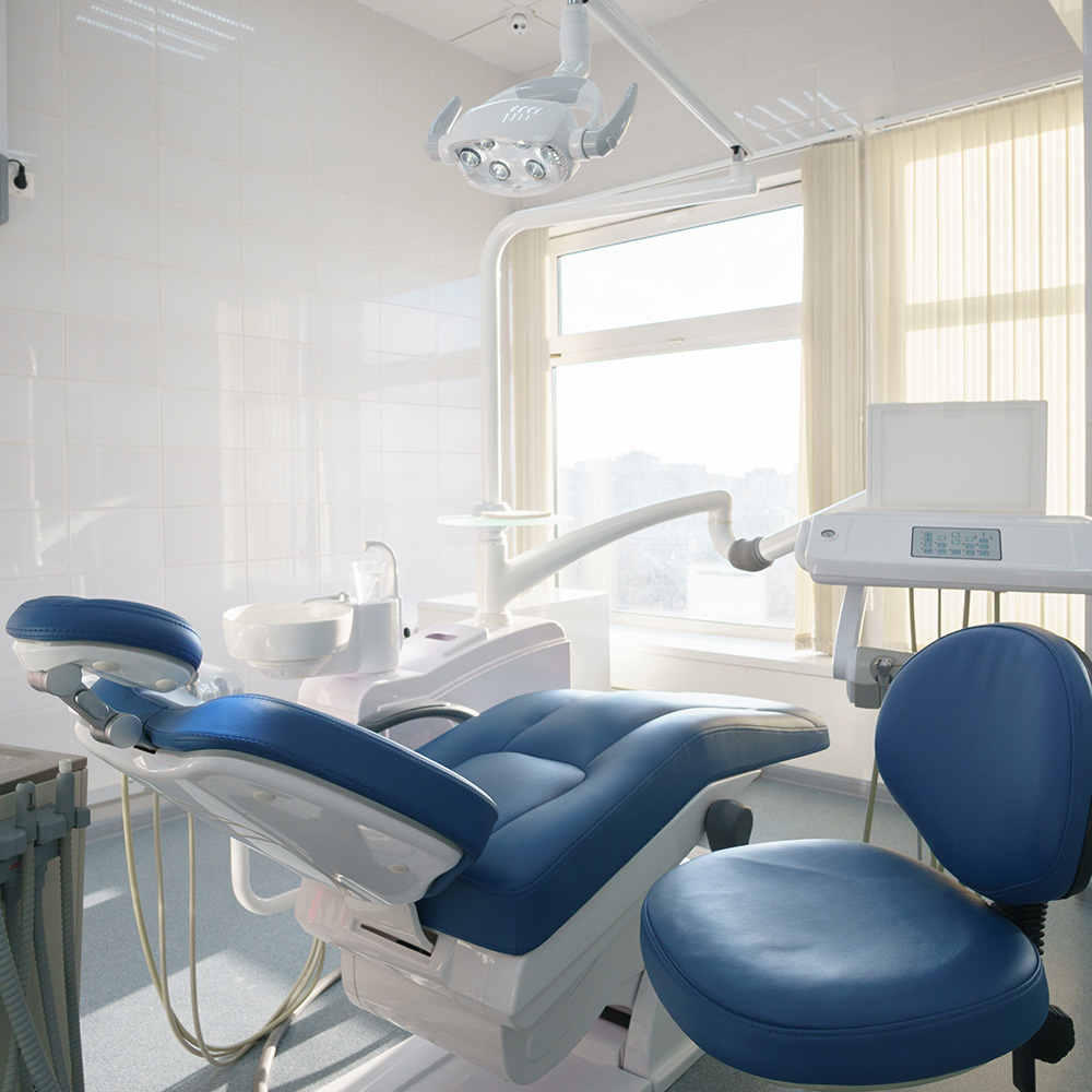
The scanning appointment begins with a brief intraoral exam and gentle retraction of the cheeks and lips to give the scanner an unobstructed view. A hygienic, single-use sleeve covers the scanner tip; no messy materials are placed in the mouth. The clinician moves the wand in a systematic pattern to capture occlusal surfaces, interproximal contacts, and soft tissue landmarks. The entire process typically takes only a few minutes per arch.
As images are captured, specialized software assembles them into a continuous 3D model. The clinician can immediately inspect the model on a monitor, zooming in to evaluate margins, contact points, and occlusion. If the scan reveals a discrepancy, targeted rescanning is quick and localized—there’s no need to repeat a full traditional impression.
Once the scan meets clinical standards, the digital file is exported in an industry-standard format and transmitted electronically to a dental laboratory or to an in-office milling unit. This direct digital transfer eliminates shipping delays and reduces the potential for deformation that can occur when physical impressions are handled, poured into stone, and shipped.
One of the most immediately noticeable advantages of digital impressions is patient comfort. Without impression trays and viscous material, there’s no gagging, fewer unpleasant tastes, and a less claustrophobic experience. This makes digital scanning particularly valuable for patients with strong gag reflexes, pronounced dental anxiety, or sensitive mouths.
Beyond comfort, digital impressions can streamline treatment timelines. Electronic transmission to a laboratory shortens turnaround times and supports more efficient scheduling. When combined with in-office CAD/CAM systems, scans can be turned into restorations within a single visit, reducing the need for temporary crowns and multiple appointments in many cases.
From a record-keeping standpoint, digital files are durable and searchable. They can be archived for future reference, compared over time to monitor wear or movement, and easily shared with specialists when multidisciplinary care is required. That portability enhances continuity of care and simplifies collaboration.
Digital scanning captures high-resolution data that helps clinicians design restorations with better marginal fit and occlusal harmony. The software’s ability to analyze and visualize the dentition in three dimensions allows for more precise planning of crown margins, implant positions, and interproximal contacts. That precision often translates into fewer adjustments at delivery and a lower incidence of remakes.
Integrated workflows—where a scan is combined with digital bite records and, when needed, CBCT data—enable more predictable restorative and implant outcomes. For implant cases, the combination of 3D surface scans and volumetric imaging supports guided workflows that improve implant placement accuracy and simplify the design of final restorations.
Digital impressions also facilitate improved communication with dental laboratories. Technicians receive clear, standardized files and can work within a digital environment to assess and refine restorations before fabrication. This reduces misunderstandings and helps ensure the final prosthetic matches the clinician’s intent.
No special preparation is required for most digital scans. Patients should maintain normal oral hygiene and remove removable appliances if instructed. During the scan, minimal cooperation is needed—simply follow the clinician’s guidance, keep the mouth relaxed, and occasionally reposition as requested to allow access to posterior areas.
After the scan, there is typically no recovery period. If your care involves a same-day restoration, the clinician will explain the sequence for milling and finishing. For laboratory-fabricated work, the digital file will be sent electronically and you’ll return for delivery when the restoration is complete. Throughout the process, the team will review the planned timeline and set expectations so you remain informed every step of the way.
Digital impressions are non-invasive and safe for virtually all patients. They are compatible with other advanced technologies used in modern dentistry, supporting an efficient, evidence-based approach to restorative care that emphasizes comfort, accuracy, and predictability.
Digital impressions represent a modern, patient-friendly way to capture the details of your smile. By replacing impression materials with precise optical scanning, this approach improves comfort, shortens turnaround times, and supports highly accurate restorative and implant workflows. The result is a smoother clinical experience and better-coordinated care.
If you’d like to learn how digital impressions are used in our Sugar Land practice or whether they’re appropriate for your upcoming treatment, please contact Dentistry of Sugar Land for more information. Our team can explain the process, answer questions, and help you prepare for your visit.


Digital impressions use an intraoral scanner to capture a precise three-dimensional image of the teeth and surrounding oral tissues. A small wand acquires hundreds or thousands of images that specialized software stitches into a single, accurate model. The resulting digital file can be reviewed from multiple angles, measured to fine tolerances, and stored in the patient record for future reference.
This data-driven workflow replaces traditional putty and trays and reduces material-related variables and handling errors. Clinicians gain immediate visual feedback and the ability to rescan locally when needed, which improves predictability for restorations. Patients benefit from increased comfort and a faster transition from examination to treatment planning.
The scanning appointment begins with a brief intraoral exam and retraction of cheeks and lips to provide an unobstructed view. A hygienic single-use sleeve covers the scanner tip while the clinician moves the wand in a systematic pattern to capture occlusal surfaces, interproximal contacts, and soft-tissue landmarks. Specialized software assembles the images in real time so the clinician can inspect margins, contacts, and occlusion on a monitor.
If the model shows a discrepancy the clinician can perform a focused rescan rather than repeating an entire traditional impression. Once the scan meets clinical standards the digital file is exported in an industry-standard format for laboratory fabrication or in-office milling. Direct digital transfer reduces deformation and avoids shipping delays associated with physical impressions.
Most patients find digital scanning more comfortable because there are no bulky trays or viscous impression materials placed in the mouth. The absence of impression material reduces gagging, unpleasant tastes, and the claustrophobic feeling some people experience with traditional techniques. Scanning is noninvasive and generally causes no discomfort beyond a routine dental examination.
Digital impressions are safe for virtually all patients and are compatible with other advanced diagnostic tools used in modern dentistry. Single-use sleeves and standard infection-control protocols protect patient safety during scanning. Clinicians will advise on simple preparations, such as removing removable appliances when appropriate, to ensure an accurate scan.
Digital impressions are commonly used to fabricate crowns, bridges, inlays, onlays, and implant restorations. They also support the design of custom night guards, splints, and clear aligners for orthodontic therapy. When combined with CAD/CAM systems, scans can feed same-day restorative workflows for many clinical situations.
Beyond restoration fabrication, digital files assist with diagnostics and monitoring by allowing clinicians to compare scans over time for wear or tooth movement. Files are easily shared with specialists for coordinated care in multidisciplinary cases. The versatility of digital impressions makes them a foundation for many modern restorative and appliance workflows.
High-resolution optical scans capture detailed surface anatomy, which helps clinicians design restorations with improved marginal fit and occlusal harmony. Three-dimensional visualization allows precise planning of crown margins, interproximal contacts, and occlusion before fabrication. This precision often translates into fewer adjustments at delivery and a reduced incidence of remakes.
When scans are integrated with digital bite records and volumetric imaging clinicians can plan restorative and implant cases more predictably. Guided workflows enable more accurate implant placement and simplify the prosthetic design process. Clear digital communication with laboratories further reduces misunderstandings and streamlines final fabrication.
A straightforward scan typically takes only a few minutes per arch after the clinician completes the initial exam and soft-tissue retraction. During scanning you will be asked to relax, follow simple positioning cues, and occasionally adjust jaw or head position to give access to posterior teeth. The clinician reviews the model on-screen and can perform quick rescans of localized areas if necessary.
There is usually no recovery time after a scan and patients can resume normal activity immediately. If a same-day restoration is planned the team will explain the design, milling, and finishing sequence prior to delivery. For laboratory-fabricated work the digital file is transmitted electronically and the practice will schedule your return visit for final delivery when the restoration is complete.
Yes, surface scans from intraoral scanning can be combined with CBCT volumetric data to create a comprehensive digital plan for implant placement. This merged dataset enables the design of surgical guides that translate the prosthetic plan into precise implant positioning. Integrated planning reduces guesswork and improves coordination between the surgical and restorative phases.
After implant placement the same digital scans assist in designing final restorations that match the planned emergence profile and occlusion. Digital workflows streamline collaboration between clinicians and dental laboratories throughout implant treatment. These processes contribute to more predictable functional and esthetic outcomes for patients.
Digital scans are exported in industry-standard formats such as STL, PLY, or OBJ so laboratories can import and manipulate the models in their CAD software. Files are transmitted electronically through secure portals or encrypted transfer services to protect patient information and maintain compliance with privacy standards. Direct digital transfer reduces transit time and the risk of distortion that can occur with poured stone models.
Laboratories can review and perform digital adjustments before fabrication, which helps catch issues early and align expectations between the clinician and technician. When an office has an in-house milling unit the same file can be used to mill restorations on site for faster turnaround. Clear file standards and communication protocols make digital collaboration more efficient and reliable.
Digital scanning is highly effective but can be challenged by limited access, excessive saliva or blood, and deeply subgingival margins that hinder optical capture. In those situations clinicians may use adjunctive techniques such as retraction cord, hemostatic measures, or gingival displacement to improve visibility. If optical capture cannot meet the clinical standard a conventional impression may still be the most reliable option for that particular case.
The decision between digital and traditional impressions is based on clinical judgment and the requirements of the restoration or appliance. Many routine and complex cases are well suited to digital workflows, but clinicians will choose the method that maximizes accuracy and long-term success. Digital and conventional approaches can coexist to ensure the best outcome for each patient.
At Dentistry of Sugar Land intraoral scanning is integrated with CAD/CAM design and in-office milling to provide same-day restorations when clinically appropriate. The clinician verifies the digital design on-screen, mills the restoration using suitable materials, and performs finishing and occlusion checks before cementation or bonding. This workflow reduces the need for temporary crowns while maintaining control over fit and esthetics.
Case selection for same-day restorations depends on complexity, material needs, and the clinician's assessment of long-term prognosis. When a laboratory-fabricated solution is preferable the digital file is sent electronically and the team coordinates the delivery timeline with the patient. Our in-office digital capabilities support efficient, predictable care tailored to patient needs in Sugar Land.
Ready to schedule your next dental appointment or have questions about our services?
Contacting Dentistry of Sugar Land is easy! Our friendly staff is available to assist you with scheduling appointments, answering inquiries about treatment options, and addressing any concerns you may have. Whether you prefer to give us a call, send us an email, or fill out our convenient online contact form, we're here to help. Don't wait to take the first step towards achieving the smile of your dreams – reach out to us today and discover the difference personalized dental care can make.