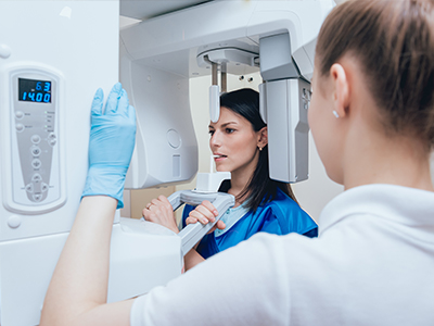
Digital radiography replaces traditional film with electronic sensors and computer processing to capture images of teeth, bones, and supporting structures. Instead of exposing film and developing it in a darkroom, a digital sensor captures the image and delivers it instantly to a computer screen. This shift from chemical-based film to a digital workflow changes how clinicians view, analyze, and store diagnostic images, making the process faster and more adaptable to modern dental care.
The advantages are practical and straightforward: images are available immediately, can be enhanced for clarity, and are stored in a patient’s digital record for long-term comparison. These capabilities help clinicians spot subtle changes over time, track healing after treatment, and collaborate more effectively with specialists when a second opinion is needed. For patients, this means a smoother visit and more informed decision-making during treatment planning.
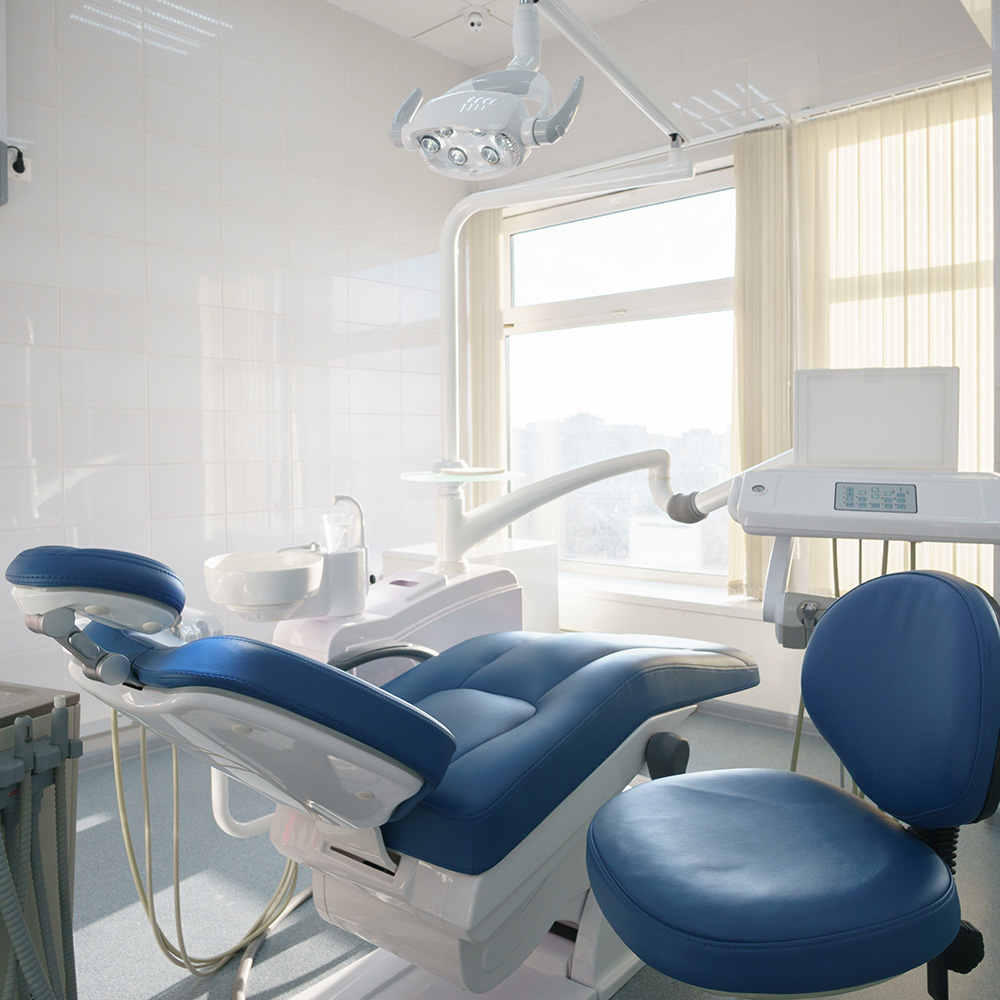
At Dentistry of Sugar Land we use digital radiography as a standard part of diagnostic care, combining precise imaging with efficient recordkeeping. The technology supports thorough evaluations while reducing the time patients spend waiting for results. Whether for routine exams or more complex diagnostic needs, digital radiography provides a dependable foundation for quality dental care.
One of the most important benefits of digital radiography is reduced radiation exposure compared with traditional film x-rays. Digital sensors are more sensitive to x‑rays, allowing clinicians to use lower doses while still producing high-quality diagnostic images. Lower exposure improves the safety profile of routine imaging, which is especially beneficial for children, pregnant patients when imaging is indicated, and patients who require frequent monitoring.
Beyond safety, speed is a major advantage. Digital images appear on the computer screen within seconds of capture, eliminating the delay associated with film processing. Instant viewing allows your dental team to assess findings right away, explain observations to you in real time, and move quickly from diagnosis to treatment planning. This responsiveness improves clinic flow and patient experience without compromising diagnostic accuracy.
Fast delivery and low exposure also support more conservative care. When clinicians can obtain clear images quickly, they’re able to detect problems earlier and recommend less invasive interventions. The combination of safety and speed makes digital radiography a practical upgrade for routine and preventive dental visits alike.
Digital radiography offers image-adjustment tools that make interpretation easier and more reliable. Brightness, contrast, and magnification can be modified without re-taking an x‑ray, helping clinicians evaluate tiny details such as hairline fractures, early decay between teeth, and the fine contours of restorative margins. These enhancements reduce the chance that important information will be missed and support more precise treatment decisions.
Advanced software also enables side‑by‑side comparisons with previous images, overlaying tools, and measurement functions that assist in planning procedures such as implant placement or endodontic assessment. When clinicians have access to sharper, manipulable images, the diagnostic process becomes both faster and more accurate, which benefits patient outcomes and long-term oral health.
Because images are digital, they can be easily anonymized and shared with specialists for collaborative review when needed. This interoperability streamlines referrals and ensures continuity of care across providers without the physical limitations of film—no envelopes, no development delays, and no loss of image fidelity in transit.
Digital radiographs are stored directly in the patient’s electronic record, creating a single, searchable repository for images, notes, and treatment histories. This integration simplifies recordkeeping and allows clinicians to view radiographs alongside clinical photos, intraoral scans, and charting during a single appointment. The result is more coherent treatment planning and better-informed conversations between the dentist and patient.
Because images are immediately accessible, coordination between hygienists, general dentists, and specialists becomes easier. For example, an intraoperative image can be reviewed instantly to confirm the position of a restoration or to guide a procedure without unnecessary interruptions. Digital storage also reduces the physical space and materials required for film archiving, supporting a more organized and environmentally conscious practice.
Integration with modern practice management systems helps protect image integrity and supports compliance with recordkeeping standards. Secure digital storage means clinicians can reliably retrieve historical images for comparison, helping to identify gradual changes early and to monitor healing after treatment over months or years.
Digital radiography improves the patient experience in several tangible ways. Sensors are typically thinner and more comfortable than traditional film packets, and the quicker imaging process reduces time spent holding uncomfortable positions. Patients also benefit from immediate, visual explanations from their dentist—the team can enlarge images, highlight areas of concern, and walk through findings together on-screen, which helps patients understand their diagnosis and treatment options.
From an environmental standpoint, digital imaging eliminates the need for chemical developers and disposable film, cutting down on hazardous waste and the materials associated with analog processing. Practices that adopt digital systems reduce both their ecological footprint and the logistical burdens tied to film handling and disposal.
Security and privacy remain priorities: digital images are managed within secure patient records, with appropriate safeguards to protect health information. When used responsibly, digital radiography delivers a blend of comfort, clarity, and sustainability that aligns with modern expectations for high-quality dental care.
Wrap-up: Digital radiography modernizes diagnosis and care by delivering faster results, clearer images, and a safer experience for patients while integrating seamlessly with today’s clinical workflows. If you’d like to learn more about how these imaging advances are used in routine exams and treatment planning, please contact us for more information.


Digital radiography uses electronic sensors and computer processing to capture images of teeth, bone, and supporting structures rather than exposing chemical film. Sensors transmit images instantly to a computer, so images are available for review within seconds instead of waiting for film development. This digital workflow changes how clinicians view, adjust, store, and compare diagnostic images.
Images can be enhanced for brightness, contrast, and magnification without re-taking an x‑ray, which improves the ability to evaluate small details. Digital files are stored in the patient record for long-term comparison and easier collaboration with specialists. At Dentistry of Sugar Land, digital radiography is used routinely to support efficient, thorough diagnostic care.
Digital images offer adjustment tools that make subtle findings easier to see, including early decay between teeth, hairline fractures, and restorative margins. Clinicians can zoom, change contrast, and apply measurements to evaluate anatomy more precisely without exposing the patient to additional radiation. These capabilities reduce the chance that clinically important details will be missed during evaluation.
Advanced software also supports side-by-side comparisons with prior images, overlay functions, and measurement aids that assist in planning complex procedures. When images can be manipulated and reviewed immediately, the diagnostic process becomes both faster and more reliable. Better visualization supports more conservative, targeted treatment decisions.
Yes, digital radiography typically requires lower radiation doses because electronic sensors are more sensitive than traditional film. Lower exposure is particularly beneficial for children, patients who need frequent monitoring, and situations where minimizing dose is important. The reduced exposure contributes to an improved safety profile for routine and repeat imaging.
Clinicians still follow established safety principles such as using shielding when appropriate and limiting imaging to clinically justified exams. Equipment calibration and proper technique ensure images are diagnostic at the lowest reasonable dose. Digital systems make it easier to capture quality images without repeat exposures, further improving safety.
During routine exams, clinicians use bitewing and periapical digital images to check for decay between teeth, assess bone levels, and evaluate the health of roots and surrounding structures. Images are captured quickly and viewed on-screen so the dentist can explain findings in real time and incorporate the images into the exam discussion. This immediacy helps patients understand their oral health and the rationale for recommended care.
Digital images also provide baseline records that clinicians can compare with future images to detect gradual changes. Hygienists and dentists can access the same images during a single appointment to coordinate care more efficiently. The streamlined process reduces appointment time spent waiting for results and supports clearer treatment planning.
Digital radiography enhances the ability to detect early-stage issues by improving image clarity and enabling image manipulation that highlights subtle changes. Early decay, small fractures, and initial bone loss are easier to spot when brightness and contrast can be adjusted and images can be magnified. Detecting problems sooner often allows for less invasive and more conservative treatment options.
Because digital files are stored and easily compared over time, clinicians can monitor progression with precise side-by-side evaluations. Regular imaging when clinically indicated helps track healing after treatment and identify gradual changes that might otherwise go unnoticed. Early detection and monitoring support better long-term oral health outcomes.
Digital radiographs are stored directly in the electronic patient record, creating a unified and searchable repository for images, notes, and treatment histories. This integration lets clinicians view radiographs alongside clinical photos, intraoral scans, and charting during a single appointment, improving coherence in treatment planning. Centralized storage also simplifies recordkeeping and reduces physical space requirements compared with film archiving.
Modern practice management systems support secure access, audit trails, and interoperability so clinicians can retrieve historical images for comparison or share them with specialists when needed. Secure storage and role-based access controls help protect patient privacy and maintain compliance with recordkeeping standards. The result is more efficient coordination of care and reliable long-term records.
Yes, digital images are typically shown to patients on a monitor during the visit so the dentist can explain findings visually and answer questions. Clinicians can enlarge areas of concern, point out early signs of decay or bone changes, and use annotations or contrast adjustments to make issues clearer. Seeing the images in real time helps patients understand the diagnosis and the options being discussed.
Clinicians strive to explain what each image shows, why a particular view was taken, and how findings relate to the proposed treatment plan. Educational discussion is part of the diagnostic process, and patients are encouraged to ask questions. When specialist input is needed, images can be reviewed collaboratively to support coordinated care.
Digital sensors are typically thinner and designed with patient comfort in mind, which often makes them more tolerable than bulky film packets. The faster capture time reduces the duration a patient needs to hold a position, an advantage for young children and patients with gag reflex sensitivity. Proper positioning aids and a gentle approach help produce diagnostic images while minimizing discomfort.
Because sensors require lower radiation doses, digital imaging is well suited for pediatric patients who may need monitoring over time. Clinicians adapt exposure settings and techniques based on patient size and clinical need to obtain diagnostic images at the lowest reasonable dose. The combined benefits of comfort and reduced exposure support safe, effective imaging for children.
Digital radiographs provide clear, measurable images that assist clinicians in evaluating anatomical landmarks, bone levels, and root morphology essential to planning implants and endodontic treatment. Measurement tools and high-resolution views help determine implant positioning and identify canal anatomy or periapical pathology. When greater three-dimensional detail is needed, digital radiography is often used alongside CBCT for comprehensive planning.
During procedures, intraoperative images can be reviewed instantly to confirm instrument position or restoration placement without unnecessary delays. The ability to compare preoperative, intraoperative, and follow-up images improves precision and helps monitor healing over time. These imaging advantages contribute to predictable treatment outcomes and coordinated care.
If you have questions about how digital radiography is used in diagnosis and treatment planning, our team is available to explain the technology, review images with you, and discuss the clinical reasons for recommended exams. A consultation allows the dentist to evaluate your history and determine the appropriate type and frequency of imaging based on your individual needs. We prioritize patient education so you can make informed decisions about your care.
To arrange an exam or speak with a member of our team about imaging protocols, please contact the practice during normal business hours and ask about diagnostic imaging services. Staff can explain what to expect during the appointment and how images will be used in your treatment plan. We aim to make the process efficient, understandable, and comfortable for every patient.
Ready to schedule your next dental appointment or have questions about our services?
Contacting Dentistry of Sugar Land is easy! Our friendly staff is available to assist you with scheduling appointments, answering inquiries about treatment options, and addressing any concerns you may have. Whether you prefer to give us a call, send us an email, or fill out our convenient online contact form, we're here to help. Don't wait to take the first step towards achieving the smile of your dreams – reach out to us today and discover the difference personalized dental care can make.