
An intraoral camera is a small, handheld imaging device designed to capture clear, full-color views of the teeth, gums, and other oral tissues. About the size of a pen, the camera uses focused lighting and a high-resolution sensor to produce detailed images that are displayed instantly on a nearby monitor. Because it offers a close-up perspective, the intraoral camera reveals surface features and soft-tissue conditions that can be difficult to see with the naked eye alone.
These cameras are engineered for maneuverability and patient comfort; their slim profiles allow clinicians to reach posterior teeth and other hard-to-see areas without causing unnecessary strain. Unlike X-rays, intraoral cameras capture surface detail in visible light rather than penetrating tissue, making them an ideal complement to radiographic imaging. The result is a fuller, more nuanced picture of oral health that helps clinicians make confident observations during an examination.
Image quality has advanced significantly in recent years, with many intraoral cameras now offering high-definition stills and video capture. This makes it possible to document subtle changes over time, assess the progress of treatment, and archive images as part of a patient’s permanent record. The immediacy and clarity of these visuals raise the standard of routine exams, allowing for earlier detection of problems that might otherwise go unnoticed.
Intraoral camera images help clinicians spot early signs of dental decay, hairline cracks, worn restorations, and areas of inflammation that may not be obvious during a visual exam alone. By enlarging the view and highlighting contrast in color and texture, the camera draws attention to potential problem areas so they can be evaluated more thoroughly. This capability supports more accurate diagnoses and reduces the risk of overlooking developing issues.
Beyond identifying existing conditions, intraoral photos contribute to thoughtful treatment planning. High-quality images allow clinicians to map the location and extent of damage, compare options for restoration, and assess the feasibility of conservative approaches versus more extensive intervention. When combined with clinical findings and radiographs, camera images help build a comprehensive treatment plan tailored to each patient’s needs.
Because these images are archived, they also serve as a visual baseline for monitoring changes over months or years. That continuity can be particularly valuable for periodontal management, tracking lesion stability, and evaluating the longevity of restorations. In short, intraoral photography enhances clinical judgment and supports more predictable patient outcomes.
One of the most powerful uses of an intraoral camera is as a communication tool. When patients can see a crisp image of their own mouth, abstract descriptions become concrete. Clinicians can point out the precise location of decay, the margins of a crack, or the inflamed tissue contributing to gum disease, which helps patients understand why a particular recommendation is being made.
This shared view fosters informed decision-making. Rather than relying solely on verbal explanations, patients see the evidence themselves and can ask targeted questions about alternatives, timing, and expected results. For many people, that visual clarity reduces anxiety and increases confidence in the recommended course of care because they better understand the condition and the rationale behind treatment choices.
Practices that use intraoral imaging tend to build stronger doctor–patient partnerships through clearer explanations and greater transparency. Showing images during the appointment creates a collaborative environment where patients feel heard and involved, which often leads to higher satisfaction with the care process.
Modern intraoral cameras integrate smoothly with digital record systems, enabling clinicians to attach images directly to a patient’s electronic chart. These photos become part of the permanent clinical record and can be referenced during future visits to track healing, evaluate treatment results, or document the progression of disease. Digital storage also simplifies retrieval and comparison of images taken at different points in time.
When collaboration with specialists or dental laboratories is required, intraoral images serve as a precise visual reference. Shared images clarify the condition and communicate important details that support coordinated care—whether the referral is for a restorative specialist, periodontist, or laboratory technician fabricating a prosthetic. Clear visuals can reduce misunderstandings and streamline the planning process.
In addition, intraoral photography complements other diagnostic technologies such as digital X-rays and cone-beam CT by providing surface-level detail that those tools do not capture. Together, these resources form a robust diagnostic toolkit that supports accurate assessments and more consistent treatment outcomes.
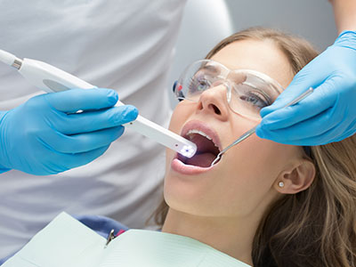
Having images taken with an intraoral camera is a quick, noninvasive part of a routine dental exam. The clinician or hygienist will gently guide the camera around the mouth while patients bite down or tilt their head as needed to expose specific areas. Sessions typically take only a few minutes, and many patients find the experience comfortable and unobtrusive.
The images are displayed in real time so the clinician can point out observations immediately and discuss next steps while the visuals are fresh. If follow-up treatment is recommended, selected images are saved to the patient’s chart so they can be reviewed later or used to track progress after the procedure. Patients are encouraged to ask questions while the images are on the screen to make sure they fully understand what the clinician sees.
Privacy and record security are handled according to standard clinical protocols; images are stored within the practice’s secure electronic record system and accessed only by authorized personnel involved in the patient’s care. Because intraoral cameras rely on visible light rather than ionizing radiation, they are safe for regular use as part of preventative and diagnostic visits.
At Dentistry of Sugar Land, we incorporate intraoral imaging into patient exams to improve clarity, communication, and the overall quality of care. If you’d like to learn more about how this technology is used during your visit, please contact us for additional information.


An intraoral camera is a small handheld imaging device that captures full-color, close-up views of the teeth, gums and other oral tissues. About the size of a pen, it uses focused lighting and a high-resolution sensor to produce detailed still images and video. The results are displayed instantly on a monitor so clinicians and patients can examine the view together.
Because it records surface detail in visible light rather than penetrating tissue, the intraoral camera is a complementary tool to radiographic imaging. Its slim, maneuverable design makes it possible to reach posterior teeth and other hard-to-see areas with minimal patient discomfort. Many cameras now support high-definition capture for documentation and comparison over time.
Intraoral images highlight color, texture and surface features that can be difficult to detect with the naked eye, such as early enamel defects, hairline cracks and worn restorations. Enlarged views make it easier for clinicians to evaluate the extent and exact location of a problem so they can form a more accurate diagnosis. This level of detail reduces the risk of overlooking developing issues and supports earlier intervention when appropriate.
High-quality photos become part of the clinical record and support more thoughtful treatment planning by documenting baseline conditions and changes over time. When used alongside clinical exams and radiographs, these images help clinicians compare restorative options and determine whether conservative measures or more extensive treatment are indicated. The visual evidence also facilitates clearer communication with specialists and laboratory technicians when coordinated care is needed.
Having images taken with an intraoral camera is a quick and noninvasive part of a routine dental exam. A clinician or hygienist will gently guide the camera around the mouth while you bite down or tilt your head as needed to expose specific areas, and the process typically takes only a few minutes. Most patients find the procedure comfortable and unobtrusive.
Images are displayed in real time so the clinician can point out observations immediately and answer questions while the visuals are fresh. Selected images are saved to the patient chart for future reference and for tracking healing or treatment progress. Privacy and record security are maintained according to standard clinical protocols.
Intraoral cameras use visible light and standard imaging sensors, so they do not expose patients to ionizing radiation and are considered safe for routine use. The procedure is noninvasive and generally causes little or no discomfort. Devices are designed for patient comfort and to minimize gagging or strain during image capture.
Clinics follow established infection control measures such as using disposable barriers or sterilizable sleeves on camera tips and following standard cleaning protocols between patients. Because the technology captures only surface-level images, it does not replace other diagnostic tools but serves as a safe, complementary method for improving visual assessment. Any specific patient concerns or sensitivities can be discussed with the clinician prior to imaging.
Modern intraoral cameras integrate with electronic record systems so images can be attached directly to a patient’s chart for easy retrieval, comparison and long-term monitoring. This digital workflow allows clinicians to review past images side by side with current photos to evaluate changes and treatment outcomes. Digital storage also streamlines collaboration with specialists and dental laboratories by providing clear visual references.
When combined with technologies such as digital X-rays and cone-beam CT, intraoral photography fills an important gap by showing surface-level detail that radiographs do not capture. Together, these tools create a robust diagnostic record that supports accurate assessments and coordinated care. Secure transmission and standardized file formats help ensure that shared images are useful to referral partners and technicians.
An intraoral camera excels at revealing surface-level signs such as early enamel changes, small cracks, marginal defects and soft-tissue inflammation that may not be obvious during a cursory visual exam. These surface findings can sometimes be identified before they become visible on radiographs, particularly when lesions are limited to enamel or the very outer tooth structure. Detecting such signs early can prompt closer monitoring or conservative intervention.
That said, intraoral imaging and radiographs are complementary; X-rays are better for seeing bone-level changes, root anatomy and interproximal decay that lie beneath the surface. Clinicians use both sources of information, together with clinical tests, to form a complete picture of oral health and to decide on the most appropriate next steps.
The frequency of intraoral imaging is determined by the clinical situation and individual patient needs rather than a fixed schedule. Many practices capture images during routine examinations, when monitoring periodontal disease, to document restorative margins, or whenever a specific concern arises. Patients with a history of dental problems or ongoing restorative work may have images taken more frequently for comparison.
Clinicians and hygienists decide when images are clinically indicated based on findings during the exam and the patient’s oral-health history. Images that document baseline conditions are useful for tracking change over months or years, and they support informed discussions about care options during the appointment. If a specialist referral or lab work is needed, additional images may be captured to facilitate clear communication.
Image quality depends on factors such as the camera’s sensor resolution, lens optics, lighting design and the operator’s technique. Good lighting and consistent focus are essential for revealing subtle color and texture differences on tooth and soft-tissue surfaces. Ergonomics and ease of positioning also influence how well the clinician can reach posterior areas and capture diagnostic views.
Software features such as autofocus, image enhancement, and the ability to capture both stills and video can improve the clinical usefulness of photos. Proper training in positioning and image capture is important to obtain consistent, interpretable images that are valuable for diagnosis and documentation. Regular maintenance and adherence to manufacturer guidance help preserve image quality over time.
Yes, intraoral images are commonly shared with specialists and dental laboratories to provide a precise visual reference for referrals and prosthetic planning. Photos clarify the condition, proportions and color relationships of teeth and soft tissues, which supports coordinated care when a specialist or technician needs to match shade, evaluate margins or plan a restoration. Clear visuals can reduce misunderstandings and speed the planning process.
Clinics transmit images securely as part of the patient’s electronic record and follow privacy practices when sharing information with outside providers. Images are used in conjunction with clinical notes, radiographs and impressions to ensure that all members of the care team have the information needed for accurate treatment planning. Patients can ask their clinician how and when their images will be shared.
Seeing a real-time image of their own mouth helps patients understand clinical findings that might otherwise be abstract or difficult to visualize. Clinicians can point to the exact location of decay, a crack or inflamed tissue and explain the reasoning behind recommendations, which supports informed decision-making. This visual approach often reduces uncertainty and helps patients ask more specific questions about alternatives and timing.
At Dentistry of Sugar Land, we use intraoral imaging to create a transparent, collaborative environment where patients feel involved in their care. Saving and reviewing images over time also helps patients see progress after treatment and reinforces the rationale for preventive strategies. Clear visuals strengthen the clinician–patient partnership and support more confident choices about oral health.
Ready to schedule your next dental appointment or have questions about our services?
Contacting Dentistry of Sugar Land is easy! Our friendly staff is available to assist you with scheduling appointments, answering inquiries about treatment options, and addressing any concerns you may have. Whether you prefer to give us a call, send us an email, or fill out our convenient online contact form, we're here to help. Don't wait to take the first step towards achieving the smile of your dreams – reach out to us today and discover the difference personalized dental care can make.

Back to top