
Oral cancer screening is a routine part of comprehensive dental care that can detect early changes in the mouth long before symptoms become obvious. Regular screening helps identify suspicious patches, lumps, or other abnormalities that may represent precancerous conditions or early-stage cancer. Early detection dramatically improves treatment options and outcomes, so understanding how screenings work and what to look for is an important part of preventive oral health.
Oral cancer refers to malignant growths that develop in the mouth and nearby structures, including the tongue, gums, floor of the mouth, lips, cheeks, and the oropharynx. These cancers can originate from the cells that line the oral cavity and can progress silently; small lesions may be present for months before they cause pain or otherwise interfere with daily life. Because some lesions are subtle, visual and tactile examination by a trained clinician is essential to spot changes that a person might otherwise miss.
Statistics show that thousands of people in the United States receive an oral cancer diagnosis each year, and a substantial number of those cases are discovered at later stages when treatment is more complex and prognoses are poorer. Detection at an early stage greatly increases the range of effective, less invasive treatments and improves long-term survival. That’s why integrating screening into routine dental checkups is considered a best practice in preventive care.
Screening is not a one-time event but an ongoing vigilance. Factors such as age, lifestyle, and emerging risk contributors mean that screening frequency should be individualized. A conversation with your dental team about personal risk and a careful, documented oral exam are the cornerstones of an effective early-detection strategy.
Traditional risk factors for oral cancer include tobacco use and heavy alcohol consumption, which together compound risk more than either factor alone. Historically, men over 50 who smoke and drink have represented a large portion of diagnosed cases, but the profile of risk is broadening. Exposure to ultraviolet (UV) light can increase risk for cancers of the lips, and environmental or chemical exposures may play a role in some cases.
Another significant and growing cause of oropharyngeal cancers is infection with certain strains of the human papillomavirus (HPV). HPV-related cancers tend to appear in different areas of the throat and often affect a younger demographic than tobacco-related cancers. While not all HPV infections lead to cancer, persistent infection with high-risk strains is associated with malignant transformation in the oropharynx.
Medical history elements such as prior radiation to the head and neck, gastroesophageal reflux disease (GERD), and nutritional deficiencies may also influence oral cancer risk. Understanding individual risk helps clinicians determine the appropriate level of surveillance and whether additional diagnostic tools should be used during screening visits.
It’s important for patients to share accurate information about lifestyle, occupational exposures, and relevant medical history so the dental team can tailor screening and education. Risk assessment is a collaborative process that supports earlier recognition and more personalized care planning.
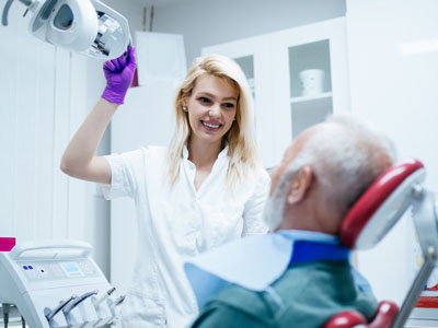
A contemporary oral cancer screening is concise, noninvasive, and typically performed as part of a routine dental exam. The clinician begins by reviewing your medical and dental history, asking about recent changes in health, persistent sore spots, difficulty swallowing, or unexplained changes in voice or oral sensation. This review provides context for the physical exam and may reveal symptoms that warrant closer inspection.
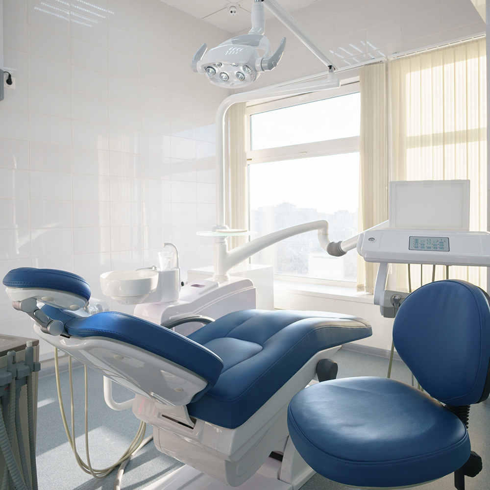
The physical portion of the screening involves a careful visual and tactile inspection of the entire oral cavity and adjacent structures. Your provider will examine the lips, tongue (top, bottom, and sides), gums, buccal mucosa (cheeks), floor of the mouth, hard and soft palate, and the oropharynx as far as is safely visible. Palpation of the neck and jawline checks for enlarged lymph nodes or other masses that can indicate deeper spread.
Some practices also use adjunctive technologies—such as specific light sources, tissue reflectance devices, or photographic documentation—to enhance visualization of suspicious areas or to track changes over time. These tools do not replace clinical judgment but can help identify lesions that merit biopsy or referral. If an area looks abnormal, the clinician will discuss next steps, which may include closer follow-up, targeted imaging, or referral to an oral surgeon or ENT specialist for definitive diagnosis.
The screening is quick and painless, usually taking only a few minutes, and helps create a baseline record for future comparisons. Documentation of findings and clear communication about any observed changes are part of routine care so patients are informed and actively involved in follow-up decisions.
Although some oral cancers are found during routine exams, patients play a critical role in early detection by reporting persistent or unusual symptoms. Warning signs include sores that do not heal within two weeks, unexplained lumps or thickening in the mouth, persistent red or white patches, and numbness or pain that isn’t attributable to an identifiable cause. Changes in speech, swallowing difficulty, a persistent sore throat, or a change in how dentures fit can also be indicators worth evaluating.
Lesions associated with oral cancer are sometimes painless in early stages, which is why visible changes should not be ignored. Any sore or discoloration that persists beyond a reasonable healing time—especially in the presence of risk factors—should prompt a professional evaluation. Patients should also note any new or persistent bleeding, dramatic changes in oral texture, or persistent ear pain that is unexplained, as these symptoms occasionally accompany oral and oropharyngeal cancers.
Keeping a personal record—photos or notes about when a lesion first appeared and whether it has changed—can be helpful when discussing symptoms with your dental team. Timely reporting and collaborative monitoring between visits make it more likely that suspicious findings are identified and managed promptly.
If a screening identifies tissue that appears suspicious, the next steps depend on the lesion’s appearance and patient risk factors. Some findings are best managed with a short-interval recheck to see if they resolve, while others require immediate referral for biopsy or imaging. A biopsy is the definitive diagnostic tool and is typically performed by an oral surgeon, ENT specialist, or another appropriate provider. Follow-up care is coordinated with the patient’s broader healthcare team to ensure timely and comprehensive evaluation.
Prevention strategies focus on minimizing known risks and maintaining a healthy oral environment. Tobacco cessation, moderating alcohol intake, protecting lips from excessive sun exposure, and addressing nutritional gaps all contribute to lower risk. Vaccination against certain strains of HPV is an important public-health tool that can reduce the incidence of HPV-related oropharyngeal cancers and is most effective when administered according to public-health guidelines.
Routine dental care, including professional cleanings and regular exams, supports early detection and overall oral health. Your dental team can provide personalized guidance about prevention, risk reduction, and the appropriate screening interval based on your health history and lifestyle. Open communication and shared decision-making help ensure that follow-up is timely and aligned with best-practice recommendations.
At Dentistry of Sugar Land, our approach to oral cancer screening emphasizes thorough exams, clear explanations, and coordinated follow-up when needed. We aim to make screening a seamless part of preventive care, giving patients confidence that changes will be recognized early and managed responsibly.
In summary, oral cancer screening is a simple yet powerful step in preserving long-term oral and overall health. Regular exams, awareness of risk factors and symptoms, and prompt follow-up when abnormalities appear are key components of effective prevention and early detection. If you have questions about screening or would like to learn more about what to expect, contact us for more information.


An oral cancer screening is a focused clinical exam designed to detect early changes in the mouth and nearby structures before symptoms become obvious. The clinician inspects and palpates the lips, tongue, gums, cheeks, floor of the mouth, palate and neck to identify suspicious patches, lumps or other abnormalities. Early detection increases treatment options and improves long-term outcomes by identifying disease at a more treatable stage.
Because many early lesions are painless and subtle, routine screening as part of a dental exam provides a baseline for future comparisons and helps catch changes that patients might not notice. A documented screening creates a record that supports timely follow-up and coordinated care if abnormalities appear. Regular screening is therefore a practical and effective component of preventive oral health.
Adults of all ages benefit from periodic oral cancer screening, and some people warrant more frequent surveillance based on risk. Individuals who use tobacco, consume alcohol heavily, have a history of head and neck radiation, or have persistent reflux or nutritional deficiencies are at higher risk and should discuss screening intervals with their dental team. Infection with high-risk strains of human papillomavirus (HPV) is an emerging cause of oropharyngeal cancers and makes screening particularly important for some patients.
Screening is individualized, so age alone does not determine need; younger people with risk factors may require closer monitoring while older adults may be screened routinely during regular dental visits. Open communication about lifestyle and medical history helps clinicians tailor the exam and follow-up plan. Sharing accurate information with your provider ensures appropriate vigilance and early intervention when needed.
A modern screening typically begins with a review of medical and dental history and questions about recent changes such as persistent sores, swallowing difficulty or unexplained numbness. The clinician then performs a careful visual and tactile exam of the oral cavity and adjacent structures, palpating the neck and jawline to detect enlarged lymph nodes or masses. This evaluation is quick, noninvasive and usually completed as part of a routine checkup.
Findings are documented and compared with prior exams to track changes over time, and the clinician will explain any observations and recommended next steps. If an area appears suspicious, options include a short-interval recheck, adjunctive imaging or referral for biopsy and specialist evaluation. Clear communication about results and follow-up expectations is a standard part of the process.
Tobacco and heavy alcohol use remain significant risk factors for oral cancer, and when combined they increase risk more than either factor alone. HPV, especially certain high-risk strains, is now a recognized cause of oropharyngeal cancers and often affects a different area of the throat than tobacco-related disease. Knowing which risk factors apply to a patient helps the dental team determine screening frequency and the level of scrutiny during examinations.
Patients with multiple risk factors may receive more frequent exams or closer follow-up when suspicious findings arise, while those with lower risk may follow routine screening intervals. Documenting risk factors also guides education about prevention and behavior modification. Personalized screening plans improve the likelihood of early detection and more favorable outcomes.
Some dental practices use adjunctive technologies to enhance visualization, such as specialized light sources, tissue-reflectance devices or photographic documentation to help identify or monitor suspicious areas. These tools can make subtle lesions more apparent and assist in tracking changes over time, but they do not replace the clinician’s visual and tactile assessment. Clinical judgment remains the primary determinant of whether further investigation is needed.
Adjunctive tests may prompt early referral or biopsy when they reveal areas of concern, and photographic records can be valuable for comparison at future visits. Patients should understand that these technologies are aids to detection rather than definitive diagnostic tools. When an area looks abnormal, a biopsy performed by an appropriate specialist is the definitive method for diagnosis.
You should schedule an evaluation if you notice any sore, lump or area of thickening that does not heal within two weeks, persistent red or white patches, or unexplained numbness or pain in the mouth. Additional warning signs include difficulty swallowing, a persistent sore throat, changes in speech or a change in how dentures fit without an obvious cause. Early-stage lesions are often painless, so visible or persistent changes warrant professional assessment.
Keeping a record of when a change first appeared and whether it has grown or altered in color or texture can be helpful when you report symptoms to your dental team. Timely reporting increases the likelihood that suspicious findings will be evaluated promptly. If you have known risk factors, erring on the side of earlier evaluation is advisable.
The frequency of screening is individualized and commonly coincides with routine dental exams, which for many adults occur every six months to a year. Patients with elevated risk factors—such as tobacco use, heavy alcohol use, prior head and neck radiation or persistent HPV infection—may require more frequent surveillance as recommended by their clinician. Your dental provider will consider your medical history, lifestyle and any prior findings when suggesting an interval.
Consistent, documented exams create a reliable baseline that makes it easier to spot subtle changes over time. Discussing your specific risk factors with your clinician ensures that your screening schedule is appropriate for your needs. At Dentistry of Sugar Land, we tailor screening intervals to individual risk and health history to support early detection.
If a clinician identifies a suspicious lesion, the immediate plan may include closer short-interval monitoring, adjunctive imaging or referral to a specialist for biopsy depending on the appearance and patient risk. A biopsy is the definitive diagnostic step and is typically performed by an oral surgeon or ENT specialist to obtain tissue for laboratory analysis. The timing and urgency of referral are based on clinical judgment and the characteristics of the lesion.
Once pathology results are available, the dental and medical teams coordinate to develop an appropriate treatment plan or further diagnostic workup as needed. Communication with the patient about findings, expected timelines and next steps is prioritized to reduce uncertainty. Coordinated care supports timely intervention and access to the right specialists when necessary.
Risk reduction focuses on addressing known contributors such as avoiding tobacco, limiting alcohol intake and protecting the lips from excessive sun exposure. Vaccination against high-risk strains of HPV is an effective public-health measure that can reduce the incidence of HPV-related oropharyngeal cancers and should be discussed with your medical provider according to current guidelines. Maintaining good nutrition and managing conditions such as chronic reflux can also help lower risk.
Regular dental care and professional cleanings support early detection and overall oral health, and clinicians can provide personalized counseling on lifestyle changes. Self-awareness and prompt reporting of persistent oral changes are practical preventive steps. Together, these measures reduce risk and improve the chance of identifying disease early when it is most treatable.
When further evaluation is indicated, the practice documents findings clearly, discusses recommended next steps with the patient, and coordinates referrals to appropriate specialists such as oral surgeons or ENT physicians. Imaging and biopsy scheduling are arranged as clinically appropriate, and the dental team communicates results and recommendations to ensure continuity of care. This coordinated approach helps patients navigate the diagnostic process with clarity and timeliness.
Patients receive explanations about what to expect at each stage and how follow-up will be managed, with an emphasis on shared decision-making and timely communication. The practice also assists in transferring records to specialists to support comprehensive evaluation and treatment planning. By coordinating care, the team seeks to make the process efficient and patient-centered.
Ready to schedule your next dental appointment or have questions about our services?
Contacting Dentistry of Sugar Land is easy! Our friendly staff is available to assist you with scheduling appointments, answering inquiries about treatment options, and addressing any concerns you may have. Whether you prefer to give us a call, send us an email, or fill out our convenient online contact form, we're here to help. Don't wait to take the first step towards achieving the smile of your dreams – reach out to us today and discover the difference personalized dental care can make.

Back to top